Overview
Diagnostic imaging describes various techniques of viewing the inside of the body to help figure out the causes of an illness or injury and confirm a diagnosis. Doctors also use it to see how well a patient’s body responds to treatment for a fracture or illness.
Diagnostic imaging allows physicians to view the inside of your body to help them find any indications of a health condition. Some machines and methods can produce pictures of the activities and structures inside your body. Your doctor will decide which medical imaging tests they’ll need to use based on the body part they’re evaluating and your symptoms.
Many imaging tests are noninvasive, easy and painless. Some will require you to remain still inside the machine for a long time, however, which can get a little uncomfortable. Some tests involve a small amount of radiation exposure.

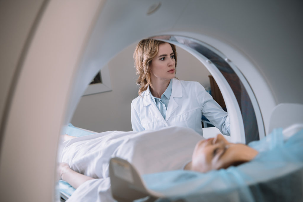
MRI
MRIs don’t use radiation, but rather a powerful magnet to obtain an image of the body of the patient. Your doctor might recommend an MRI scan for numerous reasons. It provides them with an incredibly detailed look inside your body, and they can use it to examine things like spinal cord and brain anomalies, cysts, tumors, and other bodily irregularities, breast tissue to screen for cancer, abdominal or liver diseases, and much more.
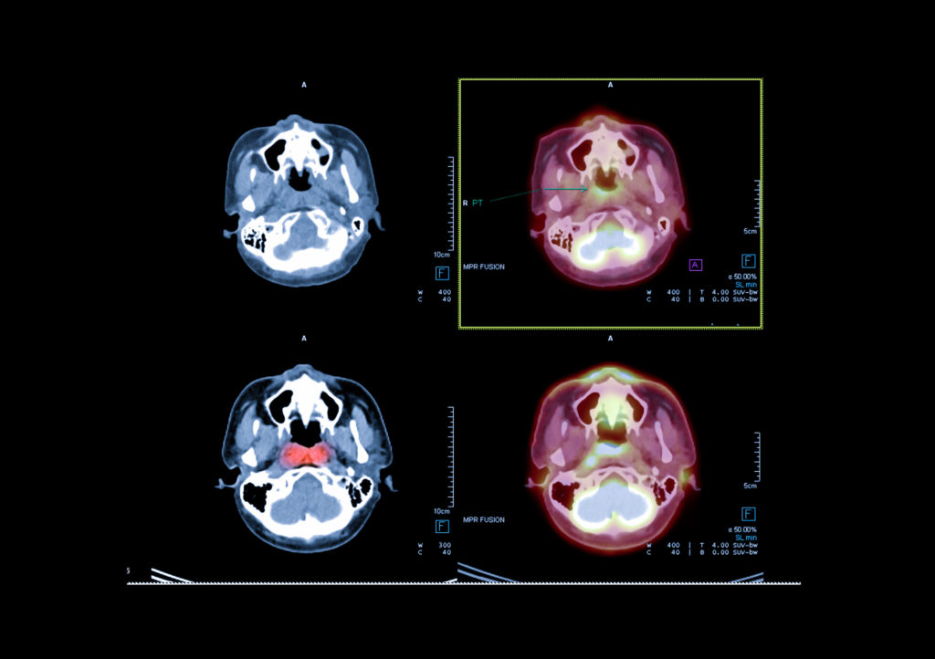
PET Scan
A PET scan, or Positron Emission Tomography scan, is a medical imaging technique used to visualize and evaluate the function and metabolism of tissues and organs in the body. It is often employed in the diagnosis, staging, and monitoring of various medical conditions, particularly cancer.
It’s important to note that PET scans provide information about the function and metabolism of tissues and organs, whereas other imaging techniques like CT (computed tomography) and MRI (magnetic resonance imaging) provide detailed anatomical images. In many cases, PET scans are combined with CT scans (PET/CT) to provide both metabolic and structural information in a single examination, enhancing the accuracy of diagnosis and treatment planning.

CT Sim
The primary purpose of a CT Sim is to precisely map the location, size, and shape of the tumor and surrounding healthy tissues within the patient’s body. This information is crucial for designing a highly targeted radiation therapy plan that maximizes the dose to the tumor while minimizing radiation exposure to healthy tissues.
Doctors can use CT scans to evaluate the spine, brain, abdomen, neck, and chest. They provide clear images of both hard and soft tissues. The pictures the CT scans produce allow doctors to quickly make medical decisions if required. Because of this quality, CT scans are commonly performed in both imaging centers and hospitals. They help physicians find injuries and diseases that could previously only be found in surgery or autopsy. While CT scans use low doses of radiation, they’re still relatively non-invasive and safe.
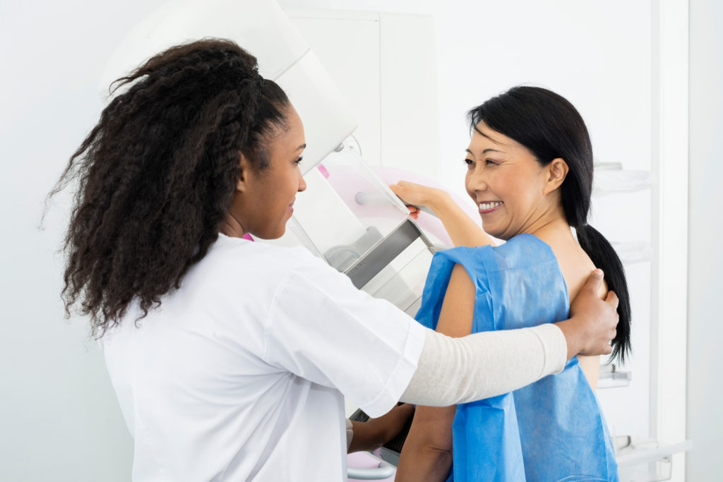
Mammogram
Mammograms are a type of x-ray image of the breasts. They check for early breast cancer signs like small lumps you or your doctor can’t feel through the use of a low-dose x-ray. Mammograms also show breast tissue changes that could be a sign of early-stage breast cancer.
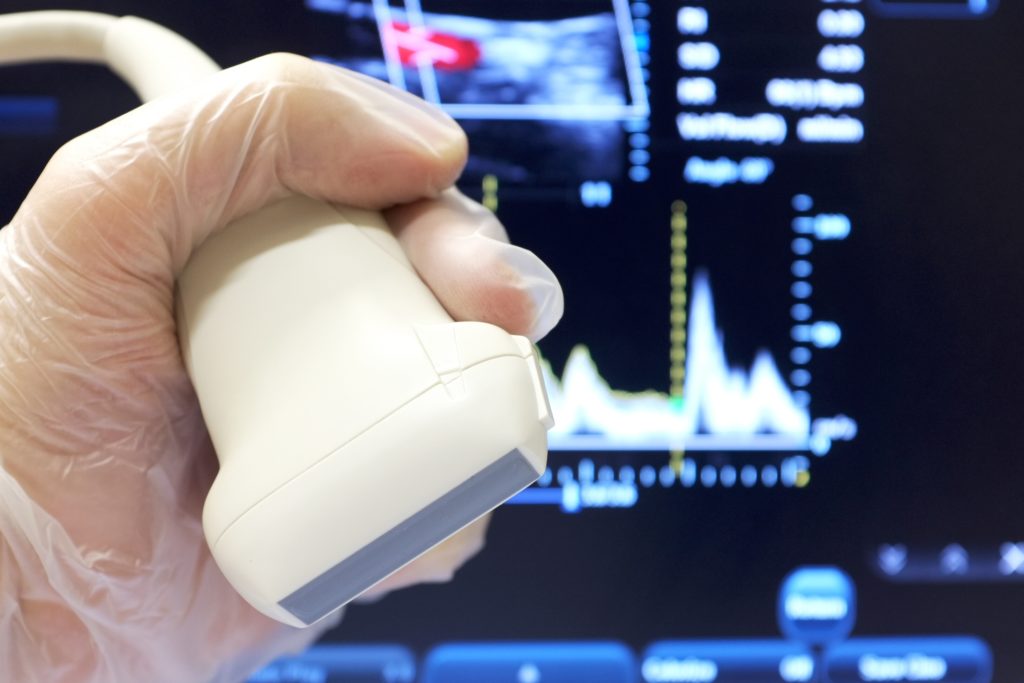
Ultrasound
Also referred to as “sonography,” ultrasound imaging is a safe imaging method that creates images of the inside of the body. It doesn’t use radiation, but rather high-frequency waves. The ultrasound images are in real-time and show the structure and movement of internal organs and the blood flow through vessels.
Doctors can diagnose a large variety of health conditions with an ultrasound. The images it creates also help physicians come up with treatment plans. If you have symptoms such as swelling, infection, or pain, your doctor might suggest an ultrasound to determine the cause. Ultrasounds are also used to assist anesthesiologists during surgical procedures when they’re guiding needles near nerves.
To learn more about our imaging partner, Premier Breast Health Institute, please CLICK HERE
Diagnostic Imaging FAQs
-
How do I prepare for diagnostic Imaging?
Please bring any previous diagnostic images as well as pathology reports to your scheduled appointment.
-
What is the purpose of diagnostic imaging?
Early and accurate diagnosis.
-
Will the exam be covered by my insurance?
Every plan is different. While most insurance plans cover radiology services there are still some that do not cover 100%. Deductibles and co-pay policies vary with different carriers. We recommend you contact your insurance company directly prior to your visit if there are any questions regarding coverage.